- บริการของเรา
- Ultrasound
อัลตราซาวนด์

- อัลตราซาวนด์ (Ultrasound)
-
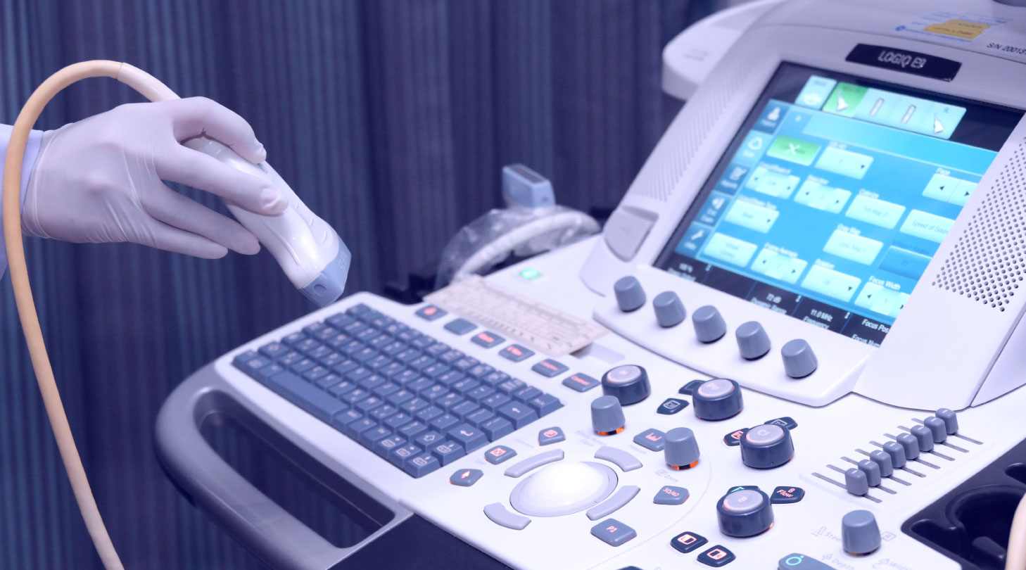
การตรวจอัลตราซาวนด์ เป็นการตรวจด้วยเครื่องมือที่ใช้คลื่นเสียงความถี่สูง โดยการส่งคลื่นเสียงความถี่สูงเข้าไป ในเนื้อเต้านม เมื่อคลื่นเสียงกระทบผ่านเนื้อเยื่อต่างๆ ภายในเต้านมจะเกิดการสะท้อนกลับของคลื่นเสียงขึ้นมาที่หัวตรวจ ซึ่งคลื่นเสียงที่ผ่านและสะท้อนกลับขึ้นมาในเนื้อเยื่อที่แตกต่างกันนี้จะถูกแปลสัญญาณโดยระบบคอมพิวเตอร์เพื่อให้ ปรากฏเป็นภาพขึ้นมา ทำให้สามารถแยกพยาธิสภาพของก้อนที่เกิดขึ้นภายในเต้านมได้ว่าเป็น ถุงน้ำ หรือ ก้อนเนื้อ และ สามารถบ่งชี้ลักษณะของก้อนเนื้องอกธรรมดา และก้อนมะเร็งได้ อีกทั้งยังช่วยระบุตำแหน่งของพยาธิสภาพที่ต้องการ นำเอาชิ้นเนื้อหรือเซลล์บริเวณนั้นออกมาวินิจฉัยเพิ่มเติมได้ โดยการใช้เข็มดูดหรือเจาะออกและส่งไปตรวจทางพยาธิวิทยา ได้อีกด้วย
การวินิจฉัยโรคมะเร็งเต้านมด้วยการตรวจอัลตราซาวนด์เพียงอย่างเดียวนั้น ไม่สามารถใช้ทดแทนการตรวจแมมโมแกรม ในการตรวจหามะเร็งเต้านมในระยะแรกเริ่มได้ เนื่องจากการตรวจด้วยเครื่องอัลตราซาวนด์ไม่สามารถตรวจพบหินปูน ขนาดเล็กที่เกิดจากมะเร็งและพยาธิสภาพอื่นๆ ได้ ดังนั้นควรตรวจอัลตราซาวนด์เต้านมร่วมกับการตรวจแมมโมแกรม เพื่อการวินิจฉัยที่ถูกต้องแม่นยำ การตรวจอัลตราซาวด์ควบคู่กับแมมโมแกรมจะช่วยเพิ่ม sensitivity และความแม่นยำ ในการวินิจฉัยมะเร็งเต้านมได้มากยิ่งขึ้น เนื่องจากลักษณะเนื้อเต้านมของผู้หญิงไทยมีความทึบรังสีมากกว่าผู้หญิง ตะวันตก การวินิจฉัยจาก Mammogram เพียงอย่างเดียวอาจจะไม่เห็นสิ่งผิดปกติทั้งหมด จึงต้องใช้ Ultrasound เข้ามาประกอบการวินิจฉัยด้วย
โดยปกติแล้ว Mammogram จะมี sensitivityในการวินิจฉัยมะเร็งที่ประมาณ 85% และจะเพิ่มสูงถึง 95% เมื่อใช้ Ultrasound มาช่วย ในปัจจุบัน ยังไม่แนะนำให้ใช้ Ultrasound แทน Mammogram ในการตรวจหามะเร็งเต้านม เพราะ Ultrasound จะไม่สามารถตรวจหาหินปูนขนาดเล็กที่ผิดปกติได้
เราจะใช้ Ultrasound ในการตรวจวินิจฉัยหลังจากการทำ Mammogram แล้ว เช่น ใช้ในการแยกถุงน้ำและก้อนเนื้อ ใช้ในผู้ที่มีเต้านมหนาทึบ เช่น อายุน้อย หญิงให้นมบุตร หรือใช้ในการทำหัตถการต่างๆ เช่น เจาะชิ้นเนื้อของเต้านม สำหรับการตรวจวินิจฉัยโรคเต้านมด้วยเครื่องอัลตราซาวนด์นั้น ทางศูนย์ถันยรักษ์จะมีรังสีแพทย์ผู้เชี่ยวชาญเฉพาะทาง เป็นผู้ทำการตรวจให้แก่ผู้มาตรวจ

